Building toward the self-driving microscope
Northwestern researcher Vinayak Dravid is integrating AI, advanced microscopy, and nanotechnology to accelerate materials discovery for energy and sustainability.
Artificial intelligence can help scientists predict promising new materials for batteries, solar panels, and other energy applications. However, predicting which materials might work is only part of the challenge. Researchers must also be able to fabricate those materials and examine them at the atomic level to understand why they perform or fail under real-world conditions. At Northwestern University, Vinayak Dravid has spent decades developing better ways to see and understand materials at the level of individual atoms. His work connects advanced microscopy, artificial intelligence, and nanotechnology to address what he calls “nanoscale solutions to gigascale challenges” in energy and sustainability.
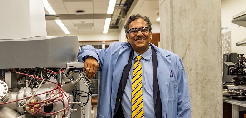
Turning microscopes into intelligent research tools
Electron microscopes are among the most powerful instruments in materials science. They can reveal atoms, defects, and chemical changes that determine whether a battery, catalyst, solar cell, or thermoelectric material will work as intended. But modern microscopes also produce enormous quantities of complex data. Reviewing those images manually is slow, and important patterns can be difficult for even experienced scientists to identify.
As the Abraham Harris Professor of Materials Science and Engineering and founding director of the Northwestern University Atomic and Nanoscale Characterization Experimental Center (NUANCE), Dravid has helped advance microscopy beyond its traditional role as a tool for producing highly magnified images. Working with experts in computer science, data science, and materials discovery, he has advanced the use of AI to interpret microscopy images, automate routine decisions, and extract information that would otherwise be difficult or time-consuming to find.
One example is a machine-learning system that Dravid and collaborators developed to classify nanoparticle images acquired through electron microscopy. The system was designed to support “megalibraries”—chips containing millions of nanoparticles with different compositions that can be screened for potentially valuable properties. Traditional characterization cannot keep pace with that volume of material. The team’s model, described in a 2024 paper in Microscopy and Microanalysis, achieved more than 95 percent precision in recognizing relevant images. The work demonstrated how AI can help automate a major bottleneck in high-throughput materials discovery.
In practical terms, this means an AI-assisted microscope could begin to make informed decisions while an experiment is underway. The instrument could recognize promising particles or unusual features and direct its attention toward them rather than collecting every possible image and leaving a researcher to sort through the results. The approach saves researchers’ time, reduces unnecessary data collection, and helps connect large-scale materials synthesis with equally rapid characterization.
Learning more while using fewer electrons
Dravid’s research also addresses a fundamental challenge in electron microscopy: the beam used to observe a material can damage or alter it. This is especially problematic for delicate materials and for experiments that follow chemical reactions as they happen.
His group has developed smart imaging approaches that ration both electron exposure and experimental time. AI and machine learning can help determine where and when the microscope should collect detailed information, allowing scientists to focus on the most valuable regions or moments rather than continuously exposing an entire sample.
This capability is important for studying materials used in energy storage, photovoltaics, catalysis, and thermoelectric devices. These materials are not static. Their atoms move, and interfaces change as the materials operate. By combining specialized experimental chambers with intelligent data collection, researchers can watch reactions and transformations at the nanoscale while reducing the risk that the measurement itself will distort the process.
The long-term goal is a closed-loop microscopy platform that can analyze incoming information, determine what and where to measure next, adjust an experiment, and pursue scientifically meaningful questions with increasing autonomy. Such a system would not replace the scientist. It would extend the scientist’s capacity by automating repetitive scanning and data analysis, enabling researchers to focus on interpreting results and designing the next generation of experiments.
Building the infrastructure for AI-enabled discovery
Dravid’s leadership extends beyond the research produced by his own laboratory. As founding director of the NUANCE Center, he has helped establish the advanced instrumentation and technical expertise needed for nanoscale research across Northwestern.
NUANCE is an interdisciplinary microscopy and surface-science facility used by hundreds of researchers from many scientific fields each year. It provides a shared environment where materials scientists, chemists, physicists, engineers, computer scientists, and external partners can work with sophisticated characterization tools and experienced technical staff.
That institutional role is particularly important for AI-driven science. Machine learning depends on abundant, reliable, and well-characterized data. In materials research, generating that data requires carefully maintained instruments, consistent experimental practices, knowledgeable staff, and collaboration between the people who understand the materials and those who develop the algorithms.
By building shared characterization infrastructure and connecting it with Northwestern’s strengths in nanotechnology, data science, and high-throughput synthesis, Dravid has helped Northwestern researchers take AI applications from isolated demonstrations to practical scientific workflows.
A bridge between AI prediction and physical discovery
Dravid’s distinctive contribution to AI for energy and sustainability lies in bringing machine intelligence into the physical laboratory. His work helps close the gap between predicting a promising material in a model and determining whether that material can perform in practice.
By integrating AI with electron microscopy, he and his collaborators are enabling faster analysis of more materials, faster recognition of important nanoscale features, and greater precision in observing dynamic processes. Through NUANCE, he has also helped provide the shared instruments, expertise, and collaborative culture required to make these approaches broadly useful.
The result is a model of AI-enabled science in which algorithms, instruments, and human judgment work together to advance energy and sustainability solutions.
Related Publications
Day, A. L., et al. “Machine Learning-Enabled Image Classification for Automated Electron Microscopy.” Microscopy and Microanalysis 30, no. 3 (2024): 456–465. https://doi.org/10.1093/mam/ozae042
Hujsak, K. A., Roth, E. W., Kellogg, W., Li, Y., and Dravid, V. P. “High Speed/Low Dose Analytical Electron Microscopy with Dynamic Sampling.” Micron 108 (2018): 31–40. https://doi.org/10.1016/j.micron.2018.03.001
Hujsak, K. A., Myers, B. D., Roth, E., Li, Y., and Dravid, V. P. “Suppressing Electron Exposure Artifacts: An Electron Scanning Paradigm with Bayesian Machine Learning.” Microscopy and Microanalysis 22, no. 4 (2016): 778–788. https://doi.org/10.1017/S1431927616011417
Wahl, C. B., Mirkin, C. A., and Dravid, V. P. “Towards Autonomous Electron Microscopy for High-Throughput Materials Discovery.” Microscopy and Microanalysis 29, supplement 1 (2023): 1913–1914. https://doi.org/10.1093/micmic/ozad067.988
Koo, K., et al. “Ultrathin Silicon Nitride Microchip for In Situ/Operando Microscopy with High Spatial Resolution and Spectral Visibility.” Science Advances 10, no. 3 (2024): eadj6417. https://doi.org/10.1126/sciadv.adj6417
Koo, K., Liu, Y., Cheng, Y., Cai, Z., Hu, X., and Dravid, V. P. “Advances and Opportunities in Closed Gas-Cell Transmission Electron Microscopy.” Chemistry of Materials 36, no. 9 (2024): 4078–4091. https://doi.org/10.1021/acs.chemmater.4c00638